- Bridging the Terahertz Gap
- Modeling the Lithium-Ion Battery
- Protection contre la Corrosion
- Modélisation des batteries
- Modélisation et Simulation dans le développement des piles à combustible
- Modélisation thermique des petits satellites
- Analyse électro-vibroacoustique d'un transducteur à armature équilibrée
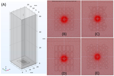
Using COMSOL to Compare the High Voltage Electric Field in Electrospinning for 3D-Printed Geometries
Electrospun nanofibers are widely used in tissue engineering as extracellular matrices (ECMs) due to their ability to support cell adhesion and direct cellular development. Fiber alignment plays a critical role in cellular behavior and is influenced by local electric field orientation during electrospinning. This study evaluates how scaffold material properties influence the electric field distribution and nanofiber alignment during electrospinning using COMSOL Multiphysics. The model employed the Electrostatics interface within the AC/DC Module and used a stationary study to simulate electric field behavior in an air-filled chamber. Scaffold geometries were imported from SolidWorks and grounded to a 6063-T83 aluminum collector plate. A steel nozzle (AISI 4340), located 60 mm above the collector, was assigned a 17 kV electric potential. Material properties for Formlabs' V4 Grey and Biomed White V1 resins were extracted from technical datasheets and manually applied to the models in COMSOL. A physics-controlled mesh with elements <0.1 mm was applied to capture field gradients near the nozzle and scaffold features. Electric field intensity was measured using line probes aligned with the x- and y-axes and visualized via MultiSlice and Streamline plots. In the V4 Grey scaffold model, the electric field strength peaked at approximately 2.2 × 10⁵ V/m along scaffold struts and dropped to 1.5 × 10⁵ V/m at the center of scaffold holes. Field vectors shifted inward toward hole centers, with square scaffold designs producing ~45° vector orientations relative to the collector normal—closely matching the nanofiber deposition angles observed under the scanning electron microscopy (SEM) images. Rectangular scaffold designs redirected the field lines perpendicular to their long axis, resulting in anisotropic fiber alignment. In contrast, the absence of a scaffold yielded a uniform electric field of approximately 1.7 × 10⁴ V/m across the probe region with vertically directed field lines. Further COMSOL simulations on the Biomed White V1 are underway, which is included to assess whether its differing dielectric constant and conductivity would influence the electric field curvature and intensity relative to V4 Grey. This material selection will also be assessed to optimize the nanofiber alignment outcomes in practice. In summary, these findings provide a predictive framework for optimizing scaffold geometry and material selection in electrospinning systems. By enabling precise, simulation-driven control over electric field orientation, this approach reduces fabrication uncertainty and accelerates the design of scaffolds tailored for targeted cellular responses in regenerative medicine.